
-
Mon-Fri: 10AM to 8PM 01722665665
-
My Account
-
-
0
Total :
₹ 0.00

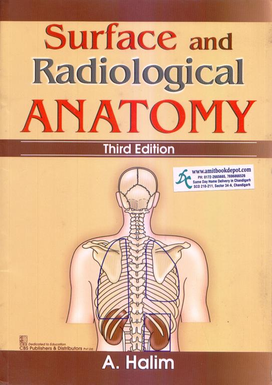
Surface and Radiological Anatomy, 3rd Edition, by A. Halim (CBS Publishers), is a practical guide for medical students to master clinically relevant anatomy. The book is split into two sections: Surface Anatomy (superior extremity, inferior extremity, thorax, abdomen and pelvis, head and neck, and brain) and Radiographic Anatomy (including bone age, vertebral column, angiography, and new imaging devices). This edition helps learners identify surface landmarks, interpret X-rays, and understand CT/MRI anatomy. Packed with diagrams and clinical pearls, it is ideal for MBBS practical exams, viva voce, and OSCE preparation. Key concepts such as palpation of pulses, lung borders, and angiographic anatomy are presented in a concise, easy-to-revise format. Essential for students of medicine, dentistry, physiotherapy, and radiology technology.


Percuss from the right 3rd intercostal space downward; liver dullness begins at the 5th rib in the midclavicular line.
The oblique fissure separates the lower lobe from the upper and middle lobes, running from T4/T5 posteriorly to the diaphragm anteriorly.
The junction of the medial one-third and lateral two-thirds of the clavicle, with needle directed toward the sternal notch.
A line drawn along the anterior humeral cortex should pass through the middle one-third of the capitellum; deviation suggests supracondylar fracture.
Feel the bony ridge extending from the zygomatic bone anteriorly to the temporal bone posteriorly, just below the lateral canthus of the eye.
Small bowel shows central valvulae conniventes spanning full width; large bowel shows haustra that do not cross the entire lumen.
Approximately 5 cm below the mastoid process along the anterior border of the trapezius muscle.
At the intersection of the midclavicular line with the 9th costal cartilage, just lateral to the rectus abdominis muscle.
Palpate the depression between the olecranon process and the coronoid process when the elbow is partially flexed.
Palpate medial to the sternocleidomastoid muscle at the level of the cricoid cartilage (C6), pressing gently backward toward the transverse processes.